Concept of tinea capitis
DOI:
https://doi.org/10.54117/gjpas.v1i2.25Keywords:
Tinea capitis, Hair follicles, Trichophyton tonsurans, Microsporum canis, Prepubertal children, TerbinafineAbstract
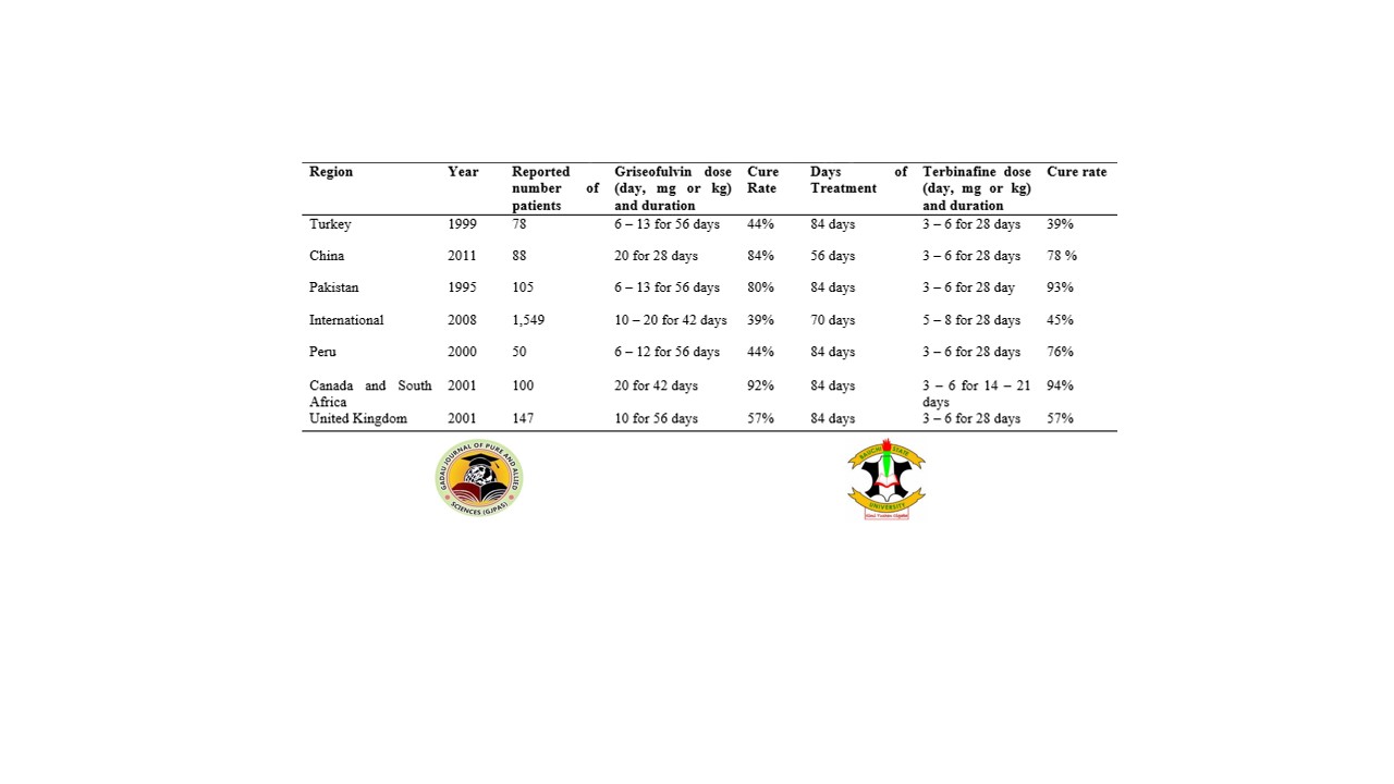
Tinea capitis is a superficial and dermatophytes infection primarily located in the hair follicles and surrounding stratum corneum. The infection is widely disseminated among prepubertal children, difficult to treat due to poor efficacy of available antifungal agents and mimic ailments related to the infection. This study reviewed the current and predominant attributes of tinea capitis including diagnostic measures and therapies against the infection. A Google search was conducted using the key term “Tinea capitis”. The search strategy includes literature reviews, clinical trials, meta-analyses, observational studies and randomized controlled trials. The result showed that Trichophyton tonsurans and Microsporum canis causes the higher number of tinea capitis cases with ectothrix and endothrix as a clinical manifestation. Terbinafine therapy had the highest cure rate in a short period of time against the infection. Non-commercial kits had the highest specificity and sensitivity than real time PCR commercial kits. The mimic ailments related to infection include trichotillomania, alopecia areata, seborrheic dermatitis, atopic dermatitis, psoriasis and bacterial scalp abscess. Allylamines and imidazole are commonly used antifungal agents against infection. Effective antifungal agents, new methods of diagnosis should be employed such as PCR targeting the 28S rDNA gene for the identification and characterization of etiological agents of tinea capitis, and practice good habits of personal and environmental hygiene should be encouraged through health education and promotion activities.
References
Abdel-Rahman, S.M., Herron, J., Fallon-Friedlander, S., Hauffe, S., Horowitz, A., Riviere, G.-J. (2005). Pharmacokinetics of Terbinafine in Young Children Treated for Tinea Capitis. The Pediatric Infectious Disease Journal 24:886–891.
Adesiji, Y.O., Omolade, B.F., Aderibigbe, I.A., Ogungbe, O.V., Adefioye, O.A., Adedokun, S.A., Adekanle, M. A., Ojedele, R. O. and Omolade, F. (2019). Prevalence of tinea capitis among children in Osogbo, Nigeria, and the associated risk factors. Diseases 7: 13.
Albengres, E., Le Louet, H. and Tillement, J.P. (1998). Systemic antifungal agents. Drug interactions of clinical significance. Drug Safety 18 (2): 83–97.
Alkeswani, A., Cantrell, W., and Elewski, B. (2019). Treatment of Tinea Capitis. Skin Appendage Disorders 5: 201 – 210.
Bennassar, A. and Grimalt, R. (2010). Management of tinea capitis in childhood. Clinical Cosmetic Investigational Dermatology 3: 89-98.
Bergman, A., Heimer, D., Kondori, N. and Enroth, H. (2013). Fast and specific dermatophyte detection by automated DNA extraction and real-time PCR. Clinical Microbiology Infection 19: E205–E211.
Caceres-Rios, H., Rueda, M., Ballona, R. and Bustamante, B. (2000). Comparison of terbinafine and griseofulvin in the treatment of tinea capitis. Journal of the American Academy of Dermatology 42: 80–4.
Chen, X., Jiang, X., Yang, M., Gonzalez, U., Lin, X., Hua, X., et al. (2016). Systemic antifungal therapy for tinea capitis in children. Cochrane Database Systematic Reviews 5: CD004685.
Coulibaly, O., Thera, M.A., Piarroux, R., Doumbo, O.K. and Ranque, S. (2015). High dermatophyte contamination levels in hairdressing salons of a West African suburban community. Mycoses 58: 65–68.
Deng, S., Hu, H., Abliz, P., Wan, Z., Wang, A., Cheng, W., et al. (2011). A random comparative study of terbinafine versus griseofulvin in patients with tinea capitis in Western China. Mycopathologia 172 (5): 365–72.
Dogo, J., Afegbua, S.L. and Dung, E.C. (2016). Prevalence of tinea capitis among school children in Nok community of Kaduna state, Nigeria. Journal of Pathogens 2016: 9601717.
Durosaro, O., Davis, M.D., Reed, K.B., et al. (2009). Incidence of cutaneous lupus erythematosus, 1965-2005: a population-based study. Archives of Dermatology 145 (3):249-253.
Elewski, B.E. (2000). Tinea capitis: a current perspective. Journal of the American Academy of Dermatology 42: 1–20.
Elewski, B.E., Caceres, H.W., Deleon, L., El-Shimy, S., Hunter, J.A., Korotkiy, N., et al. (2008). Terbinafine hydrochloride oral granules versus oral griseofulvin suspension in children with tinea capitis: results of two randomized, investigatorblinded, multicenter, international, controlled trials. Journal of the American Academy of Dermatology 59 (1): 41–54.
Ely, W.J., Rosenfeld, S. and Stones S. M. (2014) Diagnosis and management of tinea infections. American Family Physician 90 (10): 702 – 711.
Emele, F.E. and Oyeka, C.A. (2008). Tinea capitis among primary school children in Anambra state of Nigeria. Mycoses 51: 536–541.
Farooqi, M., Tabassum, S., Rizvi, D.A., Rahman, A., Rehanuddin, Awan, S., et al. (2014). Clinical types of tinea capitis and species identification in children: An experience from Tertiary Care Centres of Karachi, Pakistan. Journal of Pakistan Medical Association 64(3): 304-8.
Foster, K.W., Ghannoum, M.A. and Elewski, B.E. (2004). Epidemiologic surveillance of cutaneous fungal infection in the United States from 1999 to 2002. Journal of the American Academy of Dermatology 50 (5): 748–52.
Fuller, L.C., Smith, C.H., Cerio, R., Marsden, R.A., Midgley, G., Beard, A.L., et al. (2001) A randomized comparison of 4 weeks of terbinafine vs. 8 weeks of griseofulvin for the treatment of tinea capitis. British Journal of Dermatology 144 (2): 321–7.
GIANNI, C. (2010). Update on antifungal therapy with terbinafine. Giornale Italiano Dermatologia e Venereologia 145 (3): 415–24.
Gupta, A. K. and Summerbell, R. C. (2000). Tinea capitis. Medical Mycology 38 (4): 255–87.
Gupta, A., Alexis, M., Raboobee, N., Hofstader, S., Lynde, C., Adam, P., Summerbell, R., DE Doncker, P. (1997). Itraconazole pulse therapy is effective in the treatment of tinea capitis in children: An open multicentre study. British Journal Dermatology 137:251–254.
Gupta, A.K., Adam, P., Dlova, N., Lynde, C.W., Hofstader, S., Morar, N., et al. (2001). Therapeutic options for the treatment of tinea capitis caused by Trichophyton species: griseofulvin versus the new oral antifungal agents, terbinafine, itraconazole, and fluconazole. Pediatric Dermatology 18 (5): 433–8.
Gupta, A.K., and Cooper, E.A. (2008). Update in antifungal therapy of dermatophytosis. Mycopathologia 166:353–367.
Gupta, A.K., Foley, K.A. and Versteeg, S.G. (2017). New Antifungal Agents and New Formulations Against Dermatophytes. Mycopathologia 182 (1-2): 127–41.
Haroon, T.S., Hussain, I., Aman, S., Nagi, A., Ahmad, I., Zahid, M. and Et, A. L. (1995). A randomized doubleblind comparative study of terbinafine and griseofulvin in tinea capitis. Journal of Dermatological Treatment 6 (3): 167–9.
Hay, R. J. (2017). Tinea Capitis: current Status. Mycopathologia 182 (1-2): 87–9.
Hayette, M-P., Seidel, L., Adjetey, C., Darfouf, R., Wery, M., Boreux, R., Sacheli, R., Melin, P. and Arrese, J. (2019). Clinical evaluation of the DermaGenius®Nail real-time PCR assay for the detection of dermatophytes and Candida albicans in nails. Medical Mycology 57: 277–283.
Jartarkar, R.S., Patil, A., Goldust, Y., Cockerell, J.C., Schwartz, A.R., Grabbe, S. and Goldust, M. (2022). Pathogenesis, Immunology and management of dermatophytosis. Journal of Fungi 8 (39): 1 – 15.
John, A.M., Schwartz, R.A and Janniger, C.K. (2018). The kerion: An angry tinea capitis. International Journal of Dermatology 57(1): 3-9.
Kelly, B. P. (2012). Superficial fungal infections. Pediatric Review 33 (4): e22-e37.
Krishnan-Natesan, S. (2009). Terbinafine: a pharmacological and clinical review. Expert Opinion Pharmacotherapy 10 (16): 2723–33.
Lamisil. Package Insert: LAMISIL (Terbinafine Hydrochloride) Tablets, 250 mg Drugs@FDA: FDA Approved Drug Products 2012. (Accessed on 12 May, 2022).
Leung, C.K.A., Hon, L.K., Leong F.K., Barankin B., and Lam, M.J. (2020). Tinea capitis: An Updated Review. Recent Patents on Inflammation and Allergy Drug Discovery 14(1): 58-68.
Lipozencic, J., Skerlev, M., Orofino-Costa, R., Zaitz, V.C., Horvath, A., Chouela, E., et al. (2002). Tinea Capitis Study Group. A randomized, double-blind, parallel-group, duration-finding study of oral terbinafine and open-label, high-dose griseofulvin in children with tinea capitis due to Microsporum species. British Journal of Dermatology 146 (5): 816–23.
Memisoglu, H.R., et al. (1999). Comparative study of the efficacy and tolerability of 4 weeks of terbinafine therapy with 8 weeks of griseofulvin therapy in children with tinea capitis. Journal of Dermatological Treatment 10: 196.
Michaels, B.D. and Del Rosso, J.Q. (2012). Tinea capitis in infants: Recognition, evaluation, and management suggestions. The Journal of Clinical Aesthetic Dermatology 5(2): 49-59.
Moriarty, B., Hay, R. and Morris-Jones, R. (2012). The diagnosis and management of tinea. British Medical Journal 345: e4380.
Ndiaye, M., Sacheli, R., Diongue, K., Adjetey, C., Darfouf, R., Seck, C. M., Badiane, S. A., Diallo, A. M., Dieng, T., Hayette, M-P. & Ndiaye, D. (2022). Evaluation of the multiple Real-Time PCR Derma Genius® assay for the detection of dermatophytes in hair samples from Senegal. Journal of Fungi 8 (11): 1 – 13.
Nenoff, P., Kruger, C., Ginter-Hanselmayer, G. and Tietz, H-J. (2014). Mycology-an update. Part 1: Dermatomycoses: Causative agents, epidemiology and pathogenesis. Journal der Deutschen Dermatologischen Gesellschaft 12: 188–210.
Petinataud, D., Berger, S., Ferdynus, C., Debourgogne, A., Contet-Audonneau, N. and Machouart, M. (2016). Optimising the diagnostic strategy for onychomycosis from sample collection to FUNGAL identification evaluation of a diagnostic kit for real-time PCR. Mycoses 59: 304–311.
Rasmussen, J. E. and Ahmed, A. R. (1978). Trichophytin reactions in children with tinea capitis. Archives of Dermatological 114: 371–372.
Schafer-Korting, M., Schoellmann, C. and Korting, H.C. (2008). Fungicidal activity plus reservoir effect allow short treatment courses with terbinafine in tinea pedis. Skin Pharmacology and Physiology 21 (4): 203–10.
Schechtman, R.C., Silva, N.D., Quaresma, Bernardes, Filho, F., Buçard, A.M. and Sodre, C.T. (2015). Dermatoscopic findings as a complementary tool in the differential diagnosis of the etiological agent of tinea capitis. Anais Brasileiros de Dermatologia 90(3): S13-5.
Shah, V.P., Epstein, W.L. and Riegelman, S. (1974). Role of sweat in accumulation of orally administered griseofulvin in skin. Journal of Clinical Investigation 53 (6): 1673–8.
Tey, H.L., Tan, A.S. and Chan, Y.C. (2011). Meta-analysis of randomized, controlled trials comparing griseofulvin and terbinafine in the treatment of tinea capitis. Journal of American Academy of Dermatology 64 (4): 663–70.
Uhrlab, S., Wittig, F., Koch, D., Kruger, C., Harder, M., Gaajetaan, G., Dingemans, G. and Nenoff, P. (2019). Halten die neuen molekularen teste–microarray UND realtime-polymerasekettenreaktion–zum dermatophytennachweis das, was sie versprechen? Der Hautarzt 70: 618–626.
Veasey, J.V. and Muzy, G.S.C. (2018). Tinea capitis: Correlation of clinical presentations to agents identified in mycological culture. Anais Brasileiros de Dermatologia 93(3): 465-6.
Williams, J.V., Eichenfield, L.F., Burke, B. L., et al. (2005). Prevalence of scalp scaling in prepubertal children. Pediatrics 115 (1): e1-e6.
Wisselink, G., Van Zanten, E. and Kooistra-Smid, A. (2011). Trapped in keratin; a comparison of dermatophyte detection in nail, skin and hair samples directly from clinical samples using culture and real-time PCR. Journal of Microbiological Methods 85: 62–66.

Downloads
Published
Issue
Section
License
Copyright (c) 2022 Gadau Journal of Pure and Allied Sciences

This work is licensed under a Creative Commons Attribution 4.0 International License.

